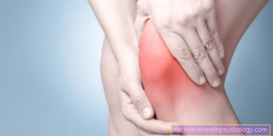
Illustratie knieholte pijn

Achterkant van de kniepijn
A - scheur van de buitenband / binnenband
B - verwondingen aan de menisci
C - artrose
D - popliteale cyste / Baker's cyste
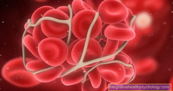
E - trombose
- Binnenmeniscus -
Mediale meniscus - Binnenband -
Ligament onderpand scheenbeen - Popliteale spier -
Popliteus spier - Scheenbeen - Scheenbeen
- Interne kuitspier -
M. gastrocnemius, caput mediale - Externe kuitspier -
M. gastrocnemius, caput laterale - Tweekoppige hamstrings
Biceps femoris spier - Halve pees spier -
Semitendinosus spier - Dijbeen - Dijbeen
- Achterste kruisband -
Achterste kruisband - Gewrichtskraakbeen -
Cartilago articularis - Voorste kruisband -
Ligament cruciatum anterius - Buitenste meniscus -
Laterale meniscus - Buitenband -
Ligamentum collaterale fibulare - Kuitbeen - Fibula
Een overzicht van alle Dr-Gumpert-afbeeldingen vindt u op: medische illustraties
Gerelateerde afbeeldingen

Illustratie
Baker's cyste

Illustratie
achterste kruisband

Illustratie
Gescheurde knieband

Illustratie
Scheur in de binnenste knieband

Illustratie
Kniegewricht

Illustratie
Knieschijf

Illustratie
Buiten kniepijn

Illustratie
Binnenkant kniepijn

Illustratie
Kraakbeenschade

Illustratie
Kruisband

Illustratie
meniscus

Illustratie
voorste kruisband